 Douleur - Allodynie/Hyperalgésie Thermique
 Douleur - Spontanée - Déficit de Posture
 Douleur - Allodynie/Hyperalgésie Mécanique
 Apprentissage/Mémoire - Attention - Addiction
 Physiologie & Recherche Respiratoire


 Douleur
 Système Nerveux Central (SNC)
  Neurodégénérescence
 Système sensoriel
 Système moteur
 Troubles de l'humeur
 Autres pathologies
 Système musculaire
 Articulations
 Métabolisme
 Thématiques transversales
 Congrès & Meetings 2026
 Authors
Wenwen Wang, Yanyan Wang, Libo Su, Mengtian Zhang, Tianyu Zhang, Jinyue Zhao, Hongyan Ma, Dongming Zhang, Fen Ji, Ryan Dingli Jiao, Hong Li, Yuming Xu, Lei Chen, Jianwei Jiao
Lab
Journal
Advanced Science
Abstract
To elucidate the molecular basis of endothelial STING deletion, we conducted RNA sequencing (RNA-seq) of WT or STING-deficient ECs in mice brains (Figure5A). The RNA-seq analysis showed that STING deletion in brain ECs induced the expression of 570 genes and reduced the expression of 635 genes (Figure5B). According to Gene Ontology (GO) analysis, the downregulated genes were enriched in terms related to cholesterol transport, cholesterol efflux, and regulation of ECs proliferation, and upregulated genes were enriched in terms related to secretion by tissue and cell-matrix adhesion (Figure5C,D). These results implied that STING might play a role in cholesterol synthesis, which plays a key role in regulating angiogenesis.[28]We selected the differential genes related to cholesterol synthesis and metabolism from the RNA-Seq of brain ECs (Figure5E) and verified the expression levels of obviously downregulated genes by RT-PCR (Figure5F). In order to further elucidate the functions of downstream genes, we constructed knockdown vectors targeting FDFT1, ABCD2, and VLDLR, which were significantly downregulated genes inSTINGECKOmice of brain ECs at E18. The results showed that the expression level of CD31 and VEGFR2 have no obvious change after the knock down of ABCD2 and VLDLR in primary brain endothelial cells (FigureS5A,B, Supporting Information), while the knockdown of FDFT1 reduced the expression level of CD31 and VEGFR2 (Figure5G), suggesting that FDFT1 has a STING KO-like phenotype. Thus, we chose FDFT1 as a promising candidate molecule. Immunofluorescent staining confirmed that FDFT1 was expressed in ECs (FigureS5C, Supporting Information). Western blotting analysis also further proved that the expression of FDFT1 was obviously reduced in knockout ECs (Figure5H,I). And we have examined the protein expression and localization of CD31 and VEGFR2 proteins after knockdown of FDFT1. The results showed that knockdown of FDFT1 reduce the expression of CD31 and VEGFR2, suggesting that FDFT1 affected angiogenesis (FigureS5D-I, Supporting Information). We then explored the regulation of FDFT1 expression by STING, and screened several candidates, including NF-ðB, TBK1, IKKð½, which can interact with STING and regulate gene expression.[29,30]We found that phosphorylated NF-ðB (p65) and phosphorylated IKKð½ were significantly downregulated, while the activation of TBK1 was not affected in ECs ofSTINGECKOmice (Figure5J,K; FigureS5J-L, Supporting Information). To validate whether NF-ðB activates FDFT1 expression, we conducted the chromatin immunoprecipitation (ChIP) experiment. The results showed that NF-ðB mainly bound to the FDFT1 promoter 1kb upstream of the transcriptional start site (Figure5L). Furthermore, we found that the binding enrichment of NF-ðB on the FDFT1 promoter reduced after the deletion of STING in ECs (Figure5M), suggesting that STING regulated FDFT1 expression through NF-ðB signaling. Further, we examined whether the induction of FDFT1 caused by STING deficiency is NFkB dependent by adding BAY11-7085 (NF-kB inhibitor) and Betulinic acid (NF-kB activator). Western blot results showed that STING promotes FDFT1 expression through the NFkB signaling pathway (FigureS5M-O, Supporting Information). FDFT1 is a critical enzyme involved in the first stage of cholesterol synthesis, which plays important roles in cell proliferation and development.[31,32]Thus, we measured the total cholesterol content of isolated brain ECs, brain, and serum inSTINGWTandSTINGECKOmice. Our results showed that the total cholesterol content was reduced in ECs and brain tissue but no obvious difference was observed between the serum ofSTINGECKOandSTINGWTmice (Figure5N; FigureS5P,Q, Supporting Information). Knockdown of FDFT1in ECs reduced the total cholesterol content in ECs (FigureS5R, Supporting Information). We found that the overexpression of FDFT1 rescued the impaired proliferation of ECs caused by STING deletion (FigureS6A-C, Supporting Information). We treated STING-deficient endothelial cells with exogenous cholesterol in vitro, and detected cell proliferation with BrdU and Ki67 immunofluorescence staining. The results showed that exogenous cholesterol treatment in STING-deficient ECs rescues endothelial cell proliferation (FigureS6D,E, Supporting Information), further indicating that STING facilitates angiogenesis by promoting cholesterol synthesis. Overall, these results suggest that the loss of endothelial STING disrupts angiogenesis by inhibiting cholesterol synthesis.
Keywords/Topics
endothelial;cells;mediated;sting;regulate;oligodendrogenesis;myelination;during;brain;development
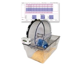
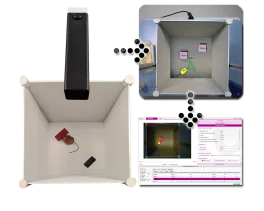
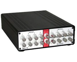
BIOSEB Instruments Used:
Grip strength test (BIO-GS4)
Source :
https://advanced.onlinelibrary.wiley.com/doi/abs/10.1002/advs.202308508