 Activité, Système Moteur & Coordination
 Douleur - Allodynie/Hyperalgésie Thermique
 Douleur - Spontanée - Déficit de Posture
 Douleur - Allodynie/Hyperalgésie Mécanique
 Anxiété & Dépression
 Apprentissage/Mémoire - Attention - Addiction
 Physiologie & Recherche Respiratoire
 Métabolisme & Nutrition




 Douleur
 Système Nerveux Central (SNC)
  Neurodégénérescence
 Système sensoriel
 Système moteur
 Troubles de l'humeur
 Autres pathologies
 Système musculaire
 Articulations
 Métabolisme
 Thématiques transversales
 Congrès & Meetings 2026
 Authors
Qi Cao, Jian Liu, Gang Huang, Su-Yuan Wang, Guo-Dong Lu, Yong Huang, Yi-Ting Chen, Zhen Zhang, Jiang-Tao Fu, Si-Jia Sun, Xiao-Fei Chen, Chunlin Zhuang, Chunquan Sheng, Fu-Ming Shen, Dong-Jie Li, Pei Wang
Lab
1; 2; 3; 1; 1; 1; 1; 4; 1; 4; 1; 1; 1; 4; 4 and
Journal
The Journal of Clinical Investigation
Abstract
We then examined muscle repair inGsdmeglobal KO (Supplemental Figure 2, A and B) and WT mice at 3 postinjury time points (D4, D7, and D14;Figure 1F). By D14, WT controls had achieved complete recovery, while KO mice continued to show multiple pathological manifestations, including reduced gastrocnemius muscle mass normalized to body weight (Figure 1G), substantial lipid accumulation in injured muscle tissue (Figure 1H), and compromised grip strength (Figure 1I). To evaluate muscle regeneration, we performed H&E staining and quantitative analysis of myofiber cross-sectional area (CSA). Histological examination revealed more severe tissue disruption in GSDME-KO mice compared with WT controls (Figure 1J). This observation was further supported by CSA quantification using the Cellpose algorithm for cellular segmentation (21), which demonstrated a significantly reduced myofiber size in GSDME-KO mice following CTX-induced injury (Figure 1J), indicating impaired regenerative capacity in the absence of GSDME. KO mice also showed diminished running distance, time, and maximum speed at D14 (Supplemental Figure 2C). Immunostaining for SDH revealed fewer SDH+fibers in KO muscle (Supplemental Figure 2D). Analysis of myosin heavy chain (MyHC) fiber types showed a consistent decrease in the ratio of oxidative type-IIa to glycolytic type-IIb fibers across time points (Supplemental Figure 2E), suggesting metabolic alterations. Seahorse assays demonstrated reduced oxygen consumption rate (OCR) and total ATP content, especially ATP from oxidative phosphorylation, in KO muscle (Supplemental Figure 2F). These findings were confirmed by luminescent ATP assays (Figure 1K). Oil Red O and perilipin-1 staining indicated increased fatty replacement in KO muscle (Figure 1LandSupplemental Figure 2G). MuSCs, marked by Pax7, are the principal cellular source for myofiber regeneration following injury. Notably, flow cytometry revealed comparable Pax7+MuSC frequencies in uninjured (D0) WT and KO mice, with both genotypes exhibiting expected activation at D4 after CTX injury but no intergenotype difference (Supplemental Figure 2H), and qPCR analysis ofPax7,Pax3, andMyf5showed unchanged expression between genotypes at D0 and equivalent upregulation at D4 (Supplemental Figure 2I), suggesting that the impaired regeneration observed in KO mice is unlikely due to early MuSCs activation defects. Collectively, these results demonstrate that GSDME deficiency markedly impairs skeletal muscle repair process, potentially due to disrupted metabolic homeostasis and lipid accumulation.
Keywords/Topics
muscle regeneration; wt mice; ko mice; grip strength; gsdme-ko mice; impaired regenerative capacity; metabolic alterations; seahorse assays; reduced oxygen consumption rate; luminescent atp assays
BIOSEB Instruments Used:
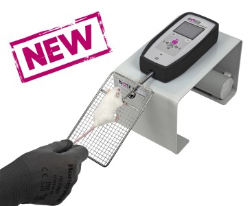
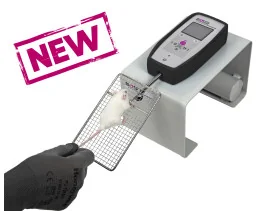
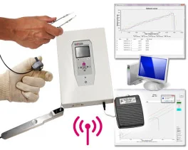
Grip strength test (BIO-GS4)
Source :