 Douleur - Allodynie/Hyperalgésie Thermique
 Douleur - Spontanée - Déficit de Posture
 Douleur - Allodynie/Hyperalgésie Mécanique
 Apprentissage/Mémoire - Attention - Addiction
 Physiologie & Recherche Respiratoire


 Douleur
 Système Nerveux Central (SNC)
  Neurodégénérescence
 Système sensoriel
 Système moteur
 Troubles de l'humeur
 Autres pathologies
 Système musculaire
 Articulations
 Métabolisme
 Thématiques transversales
 Congrès & Meetings 2026
 Authors
Yea-Eun Kim, Sang-Hyeon Hann, Young-Woo Jo, Kyusang Yoo, Ji-Hoon Kim, Jae W Lee, Young-Yun Kong
Lab
Journal
Research Square
Abstract
Freshly dissected TA or Soleus muscles were embedded in O.C.T., snap-frozen in liquid nitrogen, and stored at to80C prior to sectioning. Cross-sectional 7 m-thick sections were obtained from the embedded muscles using a cryostat. For myosin heavy chain (MyHC) staining, unfixed muscle sections were incubated overnight at 4C with mouse anti-MyHC type 1 (DSHB, BA-D5, 1:10) or mouse anti-MyHC type 2x (DSHB, 6H-1, 1:5) in addition to rat anti-laminin (Abcam, ab11576, 1:1000 dilution) in 3% BSA blocking buffer. After washes in PBS, sections were incubated for 1 h with 1:500 dilution of Alexa Fluor 488-goat anti-mouse MIgG2b (Invitrogen), or Alexa Fluor 488-conjugated anti-mouse IgM (Invitrogen), and Alexa Fluor 594-conjugated anti-rat IgG (Invitrogen). For staining myoblasts, sections were fixed in 4% paraformaldehyde (PFA) for 10 min, and washed in PBS. Antigen retrieval was then performed in citrate buffer (10 mM citric acid, pH 6) at 95C 15 min. The sections were blocked by mouse Ig blocking reagent and blocking buffer from M.O.M. Kit (Vector Laboratories), according to the manufacturer's protocol. Then, the sections were incubated with primary antibodies in the blocking buffer at 4C overnight. The primary antibodies used include mouse anti-Pax7 (1:100, DSHB), rabbit anti-Ki67 (1:500, Sigma-Aldrich), mouse anti-MyoG (1:100, DSHB), rabbit anti-Dystrophin (1:500, Abcam), rabbit anti-Laminin (1:500, Sigma-Aldrich), and rabbit anti-cleaved Caspase-3 (1:200, Cell Signaling Technologies). After washing the sections with PBS, the sections were stained with secondary antibodies for 1 hr at RT, washed, and mounted. The secondary antibodies were used at a concentration of 1:400 and include goat anti-Rabbit IgG-Alexa Fluor 488 (Thermo Fisher Scientific), goat anti-Rabbit IgG-Alexa Fluor 594 (Thermo Fisher Scientific), and goat anti-Mouse IgG-Alexa Fluor 594 (Thermo Fisher Scientific). Hoechst 33342 (1:2,000, Thermo Fisher Scientific) was used to visualize nuclei. For EdU staining, we used the Click-iT EdU Alexa Fluor 488 Imaging Kit (Thermo Fisher Scientific) following the manufacturer's protocol before the blocking step. The number of each cell type and myofibers was counted in the total TA or Soleus area, and representative images were selected in the same region of the section used in the cell counting. Imaging was conducted with EVOS FL Auto 2 (Thermo Fisher Scientific).
Keywords/Topics
Skeletal muscle ; Myofiber ; Muscle stem cells ; Myeloid / lymphoid or mixed - lineage leukemia 4
BIOSEB Instruments Used:
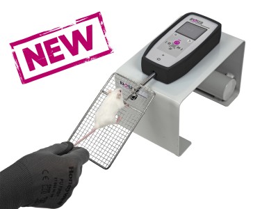
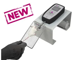
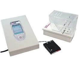
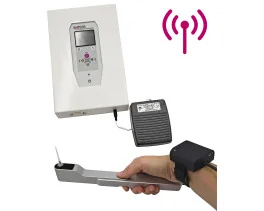
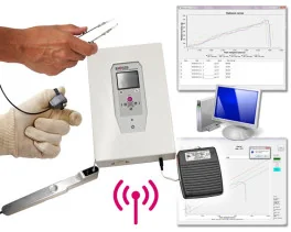
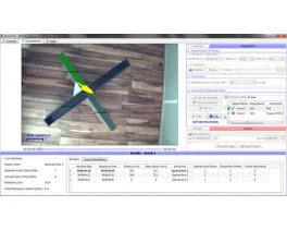
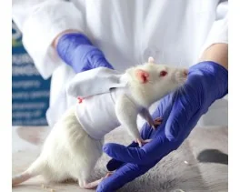
Grip strength test (BIO-GS4)
Source :