 Douleur - Allodynie/Hyperalgésie Thermique
 Douleur - Spontanée - Déficit de Posture
 Douleur - Allodynie/Hyperalgésie Mécanique
 Apprentissage/Mémoire - Attention - Addiction
 Physiologie & Recherche Respiratoire


 Douleur
 Système Nerveux Central (SNC)
  Neurodégénérescence
 Système sensoriel
 Système moteur
 Troubles de l'humeur
 Autres pathologies
 Système musculaire
 Articulations
 Métabolisme
 Thématiques transversales
 Congrès & Meetings 2026
 Authors
Dapeng Zhang, Wenzhao Wang, Shuwei Han, Huiquan Duan, Mengfan Hou, Xiaolong Zhou, Xianzheng Guo, Haosheng Chen, Xiaohong Kong, Xingshuang Zhang, Hengxing Zhou, Shiqing Feng
Lab
Journal
Theranostics
Abstract
In previous studies, researchers have used transcription factors and mRNAs to address the persistent challenge of NSC differentiation37,38. These interventions aimed to accelerate NSC differentiation and direct their fate towards becoming neurons rather than astrocytes, thereby increasing the production of neuronal cells. However, targeting these specific interventions often results in broader systemic effects. Additionally, some researchers have explored physical signals, such as electrical, mechanical, and optical signals, that influence NSC differentiation39-43. Despite these efforts, thein vivoapplication of these methods faces numerous obstacles, including uncertain outcomes, an uncontrolled intervention range, and biocompatibility issues. Moreover, some studies have failed to consider the timing and cellular environment of NSC differentiationin vivo, limiting their effectiveness. In this study, the neurotrophic factor NT3, which primarily targets NSCs while minimizing their impact on other cells, was utilized to promote NSC differentiation. Initially, the NM in the outer layer of the core-shell structure was released to reduce neuroinflammation caused by microglia, creating a favourable early-stage environment for NSC differentiation. NT3 was subsequently released to directly promote NSC differentiation at the appropriate time. The development of drug delivery systems has been a key research focus, with significant progress made in hydrogels and nanoparticles44-47. However, challenges remain48. Although hydrogels have high water content, good tissue toughness, and degradability, they often have uncontrolled drug release rates, leading to excessive local drug concentrations49,50. Nanoparticles made from metals or polymers can prolong drug action but face challenges such as being fixed in specific areas, being lost through cellular or tissue metabolism, and causing toxicity due to liver aggregation51. This study employs coaxial electrospinning technology to create a nano-fiber/net membrane delivery system. The system can locally fix and release drugs in a sustained manner through the core-shell structure of the nanofibers, addressing various aspects of SCI and enhancing therapeutic effects. However, researchers typically focus on biological properties, with the result that the mechanical properties of materials are often overlooked52,53. The dura mater surrounding the spinal cord provides essential mechanical support, and this study emphasized this aspect. Previous studies have indicated that the mechanical strength of human corpse dura mater exceeds 1 MPa, with that of mouse dura mater reaching 1.9 MPa and that of bovine dura mater reaching approximately 2 MPa54,55. The artificial dura mater developed in this study had a mechanical strength of approximately 2.2 MPa, meeting the requirements of various organisms. The addition of drugs and the use of coaxial electrospinning resulted in a nano-fiber/net structure, which was initially used in air filtration and sensor research25. This structure is an emerging direction in electrospinning, with limited biomedical research focused primarily on wound healing and drug release26,27. In this study, this structure was applied for SCI repair, improving membrane tensile strain and facilitating cell adhesion and drug release, thus expanding the scope of application.
Keywords/Topics
Spinal cord injury; coaxial electrospinning; artificial bionic dura mater; neural stem cell; nano-fiber/net
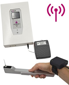
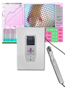
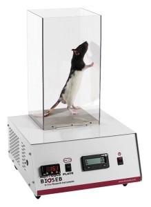
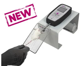
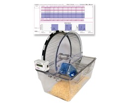
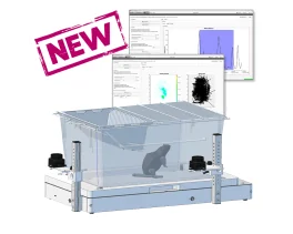
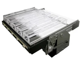
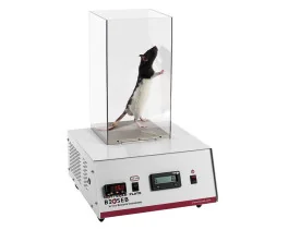
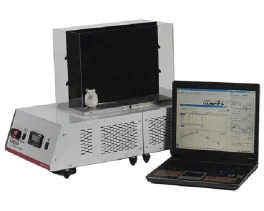
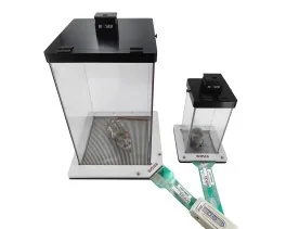
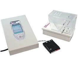
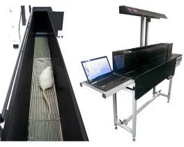
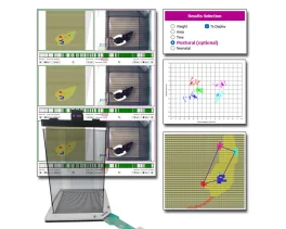
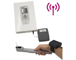
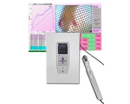
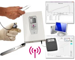
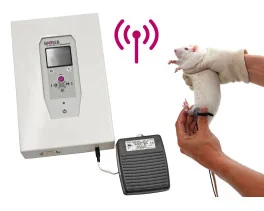
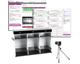
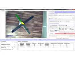
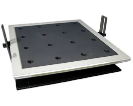
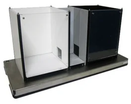
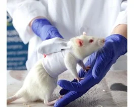
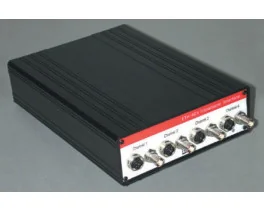
BIOSEB Instruments Used:
Cold Hot Plate Test (BIO-CHP),Electronic Von Frey - Wireless (BIO-EVF-WRS),Electronic Von Frey 5 with embedded camera (BIO-EVF5)
Source :