
 Activité, Système Moteur & Coordination
 Douleur - Allodynie/Hyperalgésie Thermique
 Douleur - Spontanée - Déficit de Posture
 Douleur - Allodynie/Hyperalgésie Mécanique
 Anxiété & Dépression
 Apprentissage/Mémoire - Attention - Addiction
 Physiologie & Recherche Respiratoire
 Métabolisme & Nutrition

















 Douleur
 Système Nerveux Central (SNC)
  Neurodégénérescence
 Système sensoriel
 Système moteur
 Troubles de l'humeur
 Autres pathologies
 Système musculaire
 Articulations
 Métabolisme
 Thématiques transversales
 Congrès & Meetings 2026
 Authors
Jun Zhang, Min Hu, Xia Wu, Mingwei Guo, Ying Ma, Jin Qiu, Siqi Wang, Yuxiang Cao, Yinzhao Zhong, Fangfang Chen, Yiwen Wang, Wei Wei, Yan Lu, Yong Zhang, Junjie Xiao, Zhenji Gan, Cheng Hu, Xinran Ma, Lingyan Xu
Lab
Journal
Advanced Science
Abstract
Next, we investigated the influences of HSF1 deficiency on the skeletal muscle of aged mice. Individual muscles exhibit distinct fiber-type compositions, i.e., the tibialis anterior (TA) is predominantly composed of fast-twitch fibers, the soleus (Sol) muscle of slow-twitch fibers, while the GAS and the quadriceps femoris (QU) muscles characterized by mixed fiber-type muscles.[24,25]22-month-old HSF1-MKO mice exhibited reduced grip strength and decreased lean mass compared with WT mice at the same age (Figure2A). These are mainly contributed by changes in fast-twitch fibers, as fast-twitch dominant (TA) and mixed-type (GAS and QU) muscles showed reduced weights, while slow-twitch dominant (Sol) muscles had comparable weights (Figure2A). RNA-seq analysis was performed in GAS muscles to understand the molecular changes in muscle after HSF1 deficiency (Figure2B). Gene ontology (GO) and KEGG analysis revealed consistent up-regulation of genes associated with autophagy and proteasomal protein catabolic process in aged HSF1-MKO mice (Figure2B,C), indicative of enhanced protein degradation, a critical event in age-related muscle atrophy,[26]which was confirmed by increased mRNA and protein levels of muscle atrophic genes (Figure2C; FigureS2A,B, Supporting Information). Besides, we observed decreased muscle hypertrophic mTOR-S6K1 signaling pathway in GAS and QU muscles of aged HSF1-MKO mice (FigureS2A,C, Supporting Information). Consistently, we found that fast-twitch fiber (TA) muscle in aged HSF1-MKO mice exhibited smaller myofiber sizes compared to aged WT mice (FigureS2D, Supporting Information), while in mixed-type muscles (GAS and QU), only fast-twitch fibers (stained positive for MYH4) were smaller in HSF1-MKO mice, with comparable slow-twitch fibers (stained positive for MYH7) (Figure2D; FigureS2E, Supporting Information). Besides, quantification of fiber-type proportions and total number of myofibers in cross-sections results demonstrated that HSF1 deficiency did not induce fiber-type switching, accompanied with the corresponding gene program (FigureS2F, Supporting Information), and not alter the total myofiber number (FigureS2G, Supporting Information). These results suggested that HSF1 deficiency mediated sarcopenia was majorly due to its regulation on the size of individual myofibers, rather than the loss of fiber numbers or fiber-type switch. Considering that loss of Pax7+satellite cells was known to attribute to sarcopenia progression, we analyzed Pax7+satellite cell numbers in aged muscle of WT and HSF1-MKO mice. The immunofluorescence data showed no significant changes in satellite cell numbers (FigureS3A, Supporting Information). It is thus possible that muscle specific deficiency of HSF1 may lead to myofiber changes, but not directly affect satellite function. However, we could not fully exclude the possibility that HSF1 mediated changes in myotubes may affect satellite cell functions via cellular crosstalk. Notably, we observed increased age-dependent occurrence of tubular aggregates as shown by prominent basophilic inclusions in muscles of aged HSF1-MKO mice (Figure2E; FigureS2H, Supporting Information).[27]Furthermore, RNA-seq analysis also revealed significant downregulation of genes involved in extracellular matrix (ECM) organization and sarcomeric component-related terms (Figure2B,F), and significant enhancement of the interferon-inflammatory signature in aged HSF1-MKO muscle (FigureS3B, Supporting Information), which was also reported previously during skeletal muscle aging.[28-32]To clarify whether the reduced ECM-related gene programs in KO mice translate into functional fibrosis benefits, we analyzed Sirius red staining, Pdgfrα+fibro-adipogenic progenitor (FAP) activity and FAP differentiation genes expression, which showed similar results between WT and HSF1-MKO mice (FigureS3C–E, Supporting Information). In contrast, muscles of HSF1-MKO mice featured decreased expressions in various collagen genes, the main components of the extracellular matrix (ECM) (Figure2F). ECM has been shown to provide critical structural support for sarcomeric anchoring and contractile function.[33]Indeed, the ultrastructure of QU muscles under TEM revealed severely abnormal sarcomeric structure, including loss of the characteristic rectangular sarcomere structure and misalignment of Z-line (Figure2G), which may contribute to muscle weakness and atrophy in aged HSF1-MKO mice. These data suggested that the fast-twitch fibers of aged HSF1-MKO mice were smaller due to enhanced protein degradation and reduced protein synthesis, which was related to muscle atrophy and disorganized muscle structure.
Keywords/Topics
aging; mitochondria; muscle atrophy; oxidative function; sarcopenia
BIOSEB Instruments Used:
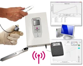
Grip strength test (BIO-GS4)
Source :
https://advanced.onlinelibrary.wiley.com/doi/abs/10.1002/advs.202510368