Authors
X Zhu, M Fréchou, M Schumacher, R Guennoun
Lab
U1195 Inserm and University Paris-Sud and University Paris-Saclay, 80 rue du Général Leclerc, 94276 Kremlin-Bicêtre, France
Journal
The Journal of Steroid BiochemistryÂ
Abstract
Treatment with progesterone limits brain damage after stroke. However, the cellular bases of the cerebroprotective effects of progesterone are not well documented. The aims of this study were to determine neural cells and functions that are affected by progesterone treatment and the role of neural progesterone receptors (PR) after stroke. Adult male PRNesCre mice, selectively lacking PR in the central nervous system, and their control PRloxP/loxP littermates were subjected to transient ischemia by middle cerebral artery occlusion (MCAO) for 30_min. Mice received either progesterone (8_mg/kg) or vehicle at 1-, 6- and 24- hrs post-MCAO and outcomes were analyzed at 48_h post-MCAO. In PRloxP/loxP mice, progesterone exerted multiple effects on different neural cell types, improved motor functional outcomes and reduced total infarct volumes. In the peri-infarct, progesterone increased the density of neurons (NeuN+ cells), of cells of the oligodendroglial lineage (Olig2+ cells) and of oligodendrocyte progenitors (OP, NG2+ cells). Progesterone decreased the density of activated astrocytes (GFAP+ cells) and reactive microglia (Iba1+ cells) coexpressing the mannose receptor type 1 CD206 marker. Progesterone also reduced the expression of aquaporin 4 (AQP4), the water channel involved in both edema formation and resorption. The beneficial effects of progesterone were not observed in PRNesCre mice. Our findings show that progesterone treatment exerts beneficial effects on neurons, oligodendroglial cells and neuroinflammatory responses via PR. These findings demonstrate that progesterone is a pleiotropic cerebroprotective agent and that neural PR represent a therapeutic target for stroke cerebroprotection.
BIOSEB Instruments Used
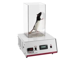
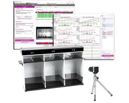
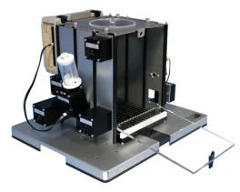
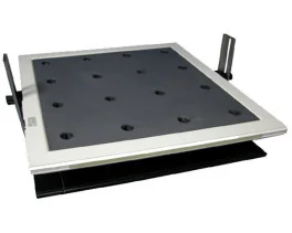
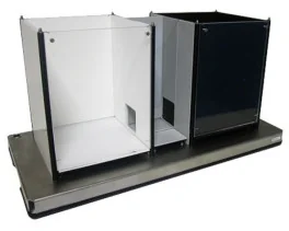
Aron Test or Four Plates Test (LE830),Rotarod (BX-ROD)
Source :
https://www.sciencedirect.com/science/article/pii/S0960076018303054

 Douleur - Allodynie/Hyperalgésie Thermique
 Douleur - Spontanée - Déficit de Posture
 Douleur - Allodynie/Hyperalgésie Mécanique
 Apprentissage/Mémoire - Attention - Addiction
 Physiologie & Recherche Respiratoire














 Douleur
 Système Nerveux Central (SNC)
  Neurodégénérescence
 Système sensoriel
 Système moteur
 Troubles de l'humeur
 Autres pathologies
 Système musculaire
 Articulations
 Métabolisme
 Thématiques transversales
 Congrès & Meetings