 Activité, Système Moteur & Coordination
 Douleur - Allodynie/Hyperalgésie Thermique
 Douleur - Spontanée - Déficit de Posture
 Douleur - Allodynie/Hyperalgésie Mécanique
 Anxiété & Dépression
 Apprentissage/Mémoire - Attention - Addiction
 Physiologie & Recherche Respiratoire
 Métabolisme & Nutrition







 Douleur
 Système Nerveux Central (SNC)
  Neurodégénérescence
 Système sensoriel
 Système moteur
 Troubles de l'humeur
 Autres pathologies
 Système musculaire
 Articulations
 Métabolisme
 Thématiques transversales
 Congrès & Meetings 2026
 Authors
Jason R Wickman, Richa Pande, Jason DaCunza, Deepa Reddy, Yuzhen Tian, Ezgi Ecem Kasimoglu, Roshell Muir, Elias K Haddad, Seena K Ajit
Lab
Journal
bioRxiv
Abstract
While our studies could not definitively observe alterations in Tregs and Th1 cells in sEV+treated animalsin vivo, we demonstrated that sEV+suppress Treg and promote Th1 polarizationin vitro. This was contrary to our initial expectations, as Treg frequency was increased in splenocyte cultures 24 hours after sEV+addition, and we detected small but potent amounts of TGF-β within sEVs. Intriguingly, the Treg population expanded in sEV+treated APC cultures showed increased T-Bet+expression, suggesting potential functional heterogeneity consistent with type 1 Treg cells[39]. However, we did not detect significant changes in complementaryin vitroassays or in ourin vivoanalyses, leaving the functional relevance of this subset unresolved. T cell polarization likely varies across anatomical sites over time as it is influenced by the location of initial antigen encounter, identity of the APC and strength of its interaction[40], as well as the local microenvironment in the tissues to which T cells subsequently migrate. Another group reported that Treg inhibit Th1 responses at the site of peripheral nerve injury to counteract neuropathic pain[24]. Future studies will need to more precisely define the timing, location, and trafficking of these different cell populations to better understand their functional contributions. Ourin vivostudies observed the strongest effect of sEV+on the increased activation of CD8+T cells in the shared CFA and sEV+draining sacral and internal iliac lymph nodes. This was accompanied by the lowest percentages of Treg and Tc1 on D1 in sEV+treated CFA animals. Previous reports have identified that CD8+T cells are necessary for the resolution of chemotherapy-induced neuropathic pain through the secretion of IL-13[25,27]. Furthermore, they showed the education of CD8+T cells by prior cisplatin exposure was necessary for mediating this resolution[26]. The importance of prior training or antigen exposure in supporting pain resolution was also demonstrated in CD4+studies mentioned above[21,22]. It is plausible that sEV+may actin vivoto prime T cells, directing them toward pro-resolving functional programs. Previous work has implicated the production of IL-13, IL-10[25,27], and enkephalins[20,22] as key mediators of these pro-resolving processes.
Keywords/Topics
dependent inflammatory pain resolution; alterations; complementaryin vitroassays; chemotherapy-induced neuropathic pain; supporting pain resolution; mechanical paw thresholds; force von frey filaments; macrophage-derived extracellular vesicles; t cell; our studies
BIOSEB Instruments Used:
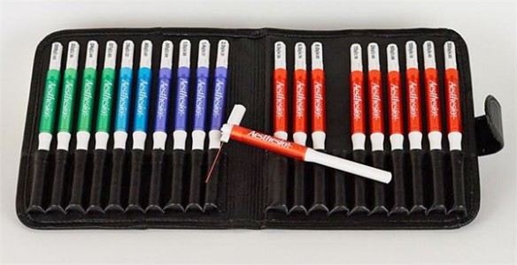
Von Frey Filaments (BIO-VF-M)
Source :