Authors
Vaysse L, Beduer A, Sol JC, Vieu C, Loubinoux I
Lab
INSERM, UMR 825 Imagerie Cérébrale et Handicaps Neurologiques, F-31024 Toulouse, France;
Journal
Biomaterials.
Abstract
With the ever increasing incidence of brain injury, developing new tissue engineering strategies to promote neural tissue regeneration is an enormous challenge. The goal of this study was to design and evaluate an implantable scaffold capable of directing neurite and axonal growth for neuronal brain tissue regeneration. We have previously shown in cell culture conditions that engineered micropatterned PDMS surface with straight microchannels allow directed neurite growth without perturbing cell differentiation and neurite outgrowth. In this study, the micropatterned PDMS device pre-seeded with hNT2 neuronal cells were implanted in rat model of primary motor cortex lesion which induced a strong motor deficit. Functional recovery was assessed by the forelimb grip strength test during 3 months post implantation. Results show a more rapid and efficient motor recovery with the hNT2 neuroimplants associated with an increase of neuronal tissue reconstruction and cell survival. This improvement is also hastened when compared to a direct cell graft of ten times more cells. Histological analyses showed that the implant remained structurally intact and we did not see any evidence of inflammatory reaction. In conclusion, PDMS bioimplants with guided neuronal cells seem to be a promising approach for supporting neural tissue reconstruction after central brain injury.
BIOSEB Instruments Used:
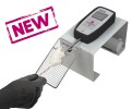
Grip strength test (BIO-GS3)

 Pain - Thermal Allodynia / Hyperalgesia
 Pain - Spontaneous Pain - Postural Deficit
 Pain - Mechanical Allodynia / Hyperalgesia
 Learning/Memory - Attention - Addiction
 Physiology & Respiratory Research
 Pain
Pain Central Nervous System (CNS)
 Neurodegeneration
 Sensory system
 Motor control
 Mood Disorders
 Other disorders
 Muscular system
 Joints
 Metabolism
 Cross-disciplinary subjects
 Preclinical studies and opioids: role in crisis management in the United States