Authors
KD Wilson, LF Ochoa, OD Solomon, R Pal, SM Cardona, VH Carpio, PH Keiser, AE Cardona, G Vargas, R Stephens
Lab
Department of Microbiology and Immunology, University of Texas Medical Branch, Galveston, TX , USA
Journal
Journal of Neuroinflammation
Abstract
Background Cerebral malaria (CM) is the most lethal outcome of Plasmodium infection. There are clear correlations between expression of inflammatory cytokines, severe coagulopathies, and mortality in human CM. However, the mechanisms intertwining the coagulation and inflammation pathways, and their roles in CM, are only beginning to be understood. In mice with T cells deficient in the regulatory cytokine IL-10 (IL-10 KO), infection with Plasmodium chabaudi leads to a hyper-inflammatory response and lethal outcome that can be prevented by anti-TNF treatment. However, inflammatory T cells are adherent within the vasculature and not present in the brain parenchyma, suggesting a novel form of cerebral inflammation. We have previously documented behavioral dysfunction and microglial activation in infected IL-10 KO animals suggestive of neurological involvement driven by inflammation. In order to understand the relationship of intravascular inflammation to parenchymal dysfunction, we studied the congestion of vessels with leukocytes and fibrin(ogen) and the relationship of glial cell activation to congested vessels in the brains of P. chabaudi-infected IL-10 KO mice.
Methods Using immunofluorescence microscopy, we describe severe thrombotic congestion in these animals. We stained for immune cell surface markers (CD45, CD11b, CD4), fibrin(ogen), microglia (Iba-1), and astrocytes (GFAP) in the brain at the peak of behavioral symptoms. Finally, we investigated the roles of inflammatory cytokine tumor necrosis factor (TNF) and coagulation on the pathology observed using neutralizing antibodies and low-molecular weight heparin to inhibit both inflammation and coagulation, respectively.
Results Many blood vessels in the brain were congested with thrombi containing adherent leukocytes, including CD4 T cells and monocytes. Despite containment of the pathogen and leukocytes within the vasculature, activated microglia and astrocytes were prevalent in the parenchyma, particularly clustered near vessels with thrombi. Neutralization of TNF, or the coagulation cascade, significantly reduced both thrombus formation and gliosis in P. chabaudi-infected IL-10 KO mice.
Conclusions These findings support the contribution of cytokines, coagulation, and leukocytes within the brain vasculature to neuropathology in malaria infection. Strikingly, localization of inflammatory leukocytes within intravascular clots suggests a mechanism for interaction between the two cascades by which cytokines drive local inflammation without considerable cellular infiltration into the brain parenchyma.
Source :
https://jneuroinflammation.biomedcentral.com/articles/10.1186/s12974-018-1207-4

 Pain - Thermal Allodynia / Hyperalgesia
 Pain - Spontaneous Pain - Postural Deficit
 Pain - Mechanical Allodynia / Hyperalgesia
 Learning/Memory - Attention - Addiction
 Physiology & Respiratory Research
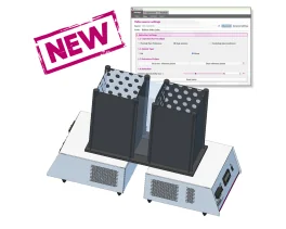
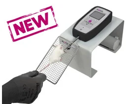
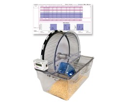

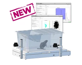
![Dynamic Weight Bearing 2.0 – Postural Module [Add-on]](https://bioseb.com/733-home_default/dynamic-weight-bearing-20-add-on-postural-module.jpg)





 Pain
Pain Central Nervous System (CNS)
 Neurodegeneration
 Sensory system
 Motor control
 Mood Disorders
 Other disorders
 Muscular system
 Joints
 Metabolism
 Cross-disciplinary subjects
 CONFERENCES & MEETINGS