 Pain - Thermal Allodynia / Hyperalgesia
 Pain - Spontaneous Pain - Postural Deficit
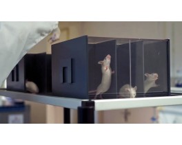
 Pain - Mechanical Allodynia / Hyperalgesia
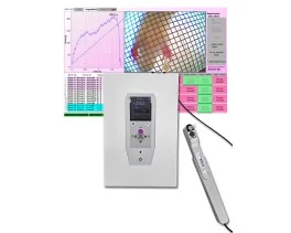
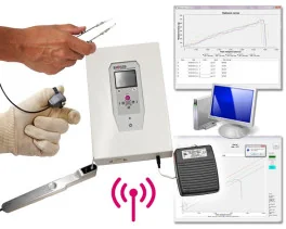
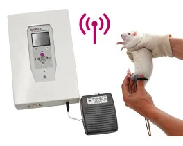
 Learning/Memory - Attention - Addiction
 Physiology & Respiratory Research
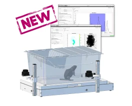

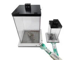
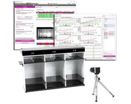
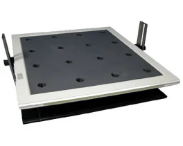
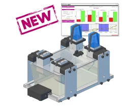
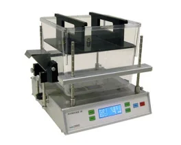
![Dynamic Weight Bearing 2.0 – Postural Module [Add-on]](https://bioseb.com/733-home_default/dynamic-weight-bearing-20-add-on-postural-module.jpg)


 Pain
Pain Central Nervous System (CNS)
 Neurodegeneration
 Sensory system
 Motor control
 Mood Disorders
 Other disorders
 Muscular system
 Joints
 Metabolism
 Cross-disciplinary subjects
 CONFERENCES & MEETINGS 2026
 Authors
D. Pinault.
Lab
Faculté de Médecine, INSERM U405, Laboratoire d’anatomo-électrophysiologie cellulaire et intégrée, Strasbourg, France.
Journal
Journal of Neuroscience Methods
Abstract
Standard large craniotomies induce undesirable brain motions during intracellular recordings in whole animal preparations. Practically all of the papers available in the literature outline a number of specific methodological approaches designed to avoid this inconvenience. Our study describes a new craniotomy–duratomy, which consists of the maintenance of a thin bone membrane and dura mater surrounding the small hole opened for lowering the recording micropipette. This new surgical preparation avoids brain movements by keeping the brain’s volume constant within the cranial cavity and does not require additional technical procedures. It is an all-purpose surgical technique, although it was developed in anaesthetized rats while studying spatio-temporal dynamics of cellular interactions associated with thalamocortical oscillations. It significantly improves both the precision of stereotaxic approaches and the success rate of single-cell recordings (e.g., current-clamp intracellular and paired recordings) compared to standard craniotomy/electrophysiology techniques.
Keywords/Topics
Miscellaneous research domains
Source :
http://www.sciencedirect.com/science/article/pii/S0165027004002419