 Pain - Thermal Allodynia / Hyperalgesia
 Pain - Spontaneous Pain - Postural Deficit
 Pain - Mechanical Allodynia / Hyperalgesia
 Learning/Memory - Attention - Addiction
 Physiology & Respiratory Research
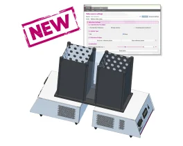

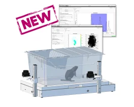

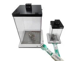
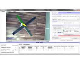
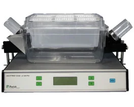
![Dynamic Weight Bearing 2.0 – Postural Module [Add-on]](https://bioseb.com/733-home_default/dynamic-weight-bearing-20-add-on-postural-module.jpg)


 Pain
Pain Central Nervous System (CNS)
 Neurodegeneration
 Sensory system
 Motor control
 Mood Disorders
 Other disorders
 Muscular system
 Joints
 Metabolism
 Cross-disciplinary subjects
 CONFERENCES & MEETINGS 2026
 Authors
Ciobanu L, Solomon E, Pyatigorskaya N, Roussel T, Le Bihan D, Frydman L
Lab
NeuroSpin, Commissariat à l'Energie Atomique et aux Energies Alternatives, Gif-sur-Yvette, France.
Journal
Neuroimage.
Abstract
This manuscript examines the origins and nature of the function-derived activation detected by magnetic resonance imaging at ultrahigh fields using different encoding methods. A series of preclinical high field (7 T) and ultra-high field (17.2 T) fMRI experiments were performed using gradient echo EPI, spin echo EPI and spatio-temporally encoded (SPEN) strategies. The dependencies of the fMRI signal change on the strength of the magnetic field and on different acquisition and sequence parameters were investigated. Artifact-free rat brain images with good resolution in all areas, as well as significant localized activation maps upon forepaw stimulation, were obtained in a single scan using fully refocused SPEN sequences devoid of T2* effects. Our results showed that, besides the normal T2-weighted BOLD contribution that arises in spin-echo sequences, fMRI SPEN signals contain a strong component caused by apparent T1-related effects, demonstrating the potential of such technique for exploring functional activation in rodents and on humans at ultrahigh fields.
Keywords/Topics
Miscellaneous research domains
Source :