 Pain - Thermal Allodynia / Hyperalgesia
 Pain - Spontaneous Pain - Postural Deficit
 Pain - Mechanical Allodynia / Hyperalgesia
 Learning/Memory - Attention - Addiction
 Physiology & Respiratory Research


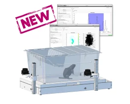


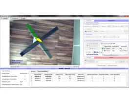
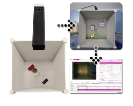
![Dynamic Weight Bearing 2.0 – Postural Module [Add-on]](https://bioseb.com/733-home_default/dynamic-weight-bearing-20-add-on-postural-module.jpg)


 Pain
Pain Central Nervous System (CNS)
 Neurodegeneration
 Sensory system
 Motor control
 Mood Disorders
 Other disorders
 Muscular system
 Joints
 Metabolism
 Cross-disciplinary subjects
 CONFERENCES & MEETINGS 2026
 Authors
M Niu, F Zhao, K Bondelid et al
Lab
Case Western Reserve University, Cleveland, OH, USA
Journal
Aging Cell
Abstract
D620N mutation in the vacuolar protein sorting 35 ortholog (VPS35) gene causes late-onset, autosomal dominant familial Parkinson's disease (PD) and contributes to idiopathic PD. However, how D620N mutation leads to PD-related deficits in vivo remains unclear. In the present study, we thoroughly characterized the biochemical, pathological, and behavioral changes of a VPS35 D620N knockin (KI) mouse model with chronic aging. We reported that this VPS35 D620N KI model recapitulated a spectrum of cardinal features of PD at 14 months of age which included age-dependent progressive motor deficits, significant changes in the levels of dopamine (DA) and DA metabolites in the striatum, and robust neurodegeneration of the DA neurons in the SNpc and DA terminals in the striatum, accompanied by increased neuroinflammation, and accumulation and aggregation of alpha-synuclein in DA neurons. Mechanistically, D620N mutation induced mitochondrial fragmentation and dysfunction in aged mice likely through enhanced VPS35-DLP1 interaction and increased turnover of mitochondrial DLP1 complexes in vivo. Finally, the VPS35 D620N KI mice displayed greater susceptibility to MPTP-mediated degeneration of nigrostriatal pathway, indicating that VPS35 D620N mutation increased vulnerability of DA neurons to environmental toxins. Overall, this VPS35 D620N KI mouse model provides a powerful tool for future disease modeling and pharmacological studies of PD. Our data support the involvement of VPS35 in the development of alpha-synuclein pathology in vivo and revealed the important role of mitochondrial fragmentation/dysfunction in the pathogenesis of VPS35 D620N mutation-associated PD in vivo.
BIOSEB Instruments Used
Grip strength test (BIO-GS3)
Keywords/Topics
Parkinson disease
Source :