Authors
A Fougerat, G Schoiswohl, A Polizzi et al
Lab
Toxalim (Research Center in Food Toxicology), INRAE, ENVT, INP- PURPAN, UMR 1331, UPS, Université de Toulouse, Toulouse, France
Journal
bioRxiv
Abstract
Objective: In hepatocytes, peroxisome proliferator-activated receptor _ (PPARalpha) acts as a lipid sensor that regulates hepatic lipid catabolism during fasting and orchestrates a genomic response required for whole-body homeostasis. This includes the biosynthesis of ketone bodies and the secretion of the starvation hormone fibroblast growth factor 21 (FGF21). Several lines of evidence suggest that adipose tissue lipolysis contributes to this specific process. However, whether adipose tissue lipolysis is a dominant signal for the extensive remodeling of liver gene expression dependent on PPARalpha has not been investigated.
Methods: First, using mice lacking adipose tissue lipolysis through adipocyte-specific deletion of adipose triglyceride lipase (ATGL), we characterized the responses dependent on adipocyte ATGL during fasting. Next, we performed liver whole genome expression analysis in fasted mice upon deletion of adipocyte ATGL or hepatocyte PPARalpha. Finally, we tested the consequences of hepatocyte-specific PPARalpha deficiency during pharmacological induction of adipocyte lipolysis with a b3-adrenergic receptor agonist.
Results: In the absence of ATGL in adipocytes, ketone body and FGF21 productions were impaired in response to starvation. Liver transcriptome analysis revealed that adipocyte ATGL is critical for regulation of hepatic gene expression during fasting and highlighted a strong enrichment in PPARalpha target genes in this condition. Genome expression analysis confirmed that a large set of fasting-induced genes are sensitive to both ATGL and PPARalpha. Adipose tissue lipolysis induced by acute activation of the b3-adrenergic receptor also triggered PPARalpha-dependent responses in the liver, supporting a role for adipocyte-derived fatty acids as dominant signals for hepatocyte PPARalpha activity. In addition, the absence of hepatocyte PPARalpha altered brown adipose tissue (BAT) morphology and reduced UCP1 expression upon stimulation of the b3- adrenergic receptor. In agreement with this finding, mice lacking hepatocyte PPARalpha showed decreased tolerance to acute cold exposure.
Conclusions These results underscore the central role of hepatocyte PPARalpha in the sensing of adipocyte-derived fatty acids and reveal that its activity is essential for full activation of BAT. Intact PPARalpha activity in hepatocytes is required for cross-talk between adipose tissues and the liver during fat mobilization during fasting and cold exposure
Source :
https://www.biorxiv.org/content/biorxiv/early/2021/01/30/2021.01.28.428684.full.pdf

 Pain - Thermal Allodynia / Hyperalgesia
 Pain - Spontaneous Pain - Postural Deficit
 Pain - Mechanical Allodynia / Hyperalgesia
 Learning/Memory - Attention - Addiction
 Physiology & Respiratory Research





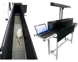
![Dynamic Weight Bearing 2.0 – Postural Module [Add-on]](https://bioseb.com/733-home_default/dynamic-weight-bearing-20-add-on-postural-module.jpg)


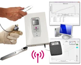
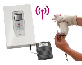


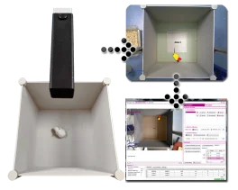









 Pain
Pain Central Nervous System (CNS)
 Neurodegeneration
 Sensory system
 Motor control
 Mood Disorders
 Other disorders
 Muscular system
 Joints
 Metabolism
 Cross-disciplinary subjects
 CONFERENCES & MEETINGS