 Pain - Thermal Allodynia / Hyperalgesia
 Pain - Spontaneous Pain - Postural Deficit
 Pain - Mechanical Allodynia / Hyperalgesia
 Learning/Memory - Attention - Addiction
 Physiology & Respiratory Research
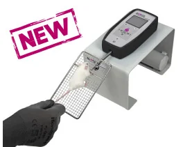

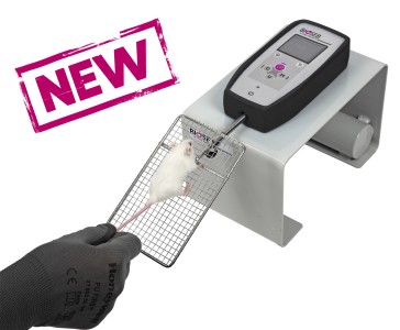
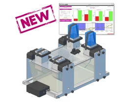
![Dynamic Weight Bearing 2.0 – Postural Module [Add-on]](https://bioseb.com/733-home_default/dynamic-weight-bearing-20-add-on-postural-module.jpg)


 Pain
Pain Central Nervous System (CNS)
 Neurodegeneration
 Sensory system
 Motor control
 Mood Disorders
 Other disorders
 Muscular system
 Joints
 Metabolism
 Cross-disciplinary subjects
 CONFERENCES & MEETINGS 2026
 Authors
Emilie Passerieux, Elodie Desplanche, Laurie Alburquerque, Quentin Wynands, Axel Bellanger, Anne Virsolvy, Fars Gouzi, Olivier Cazorla, Arnaud Bourdin, Maurice Hayot, Pascal Pomis
Lab
Journal
Acta Physiologica
Abstract
While ELA/LPS treatment of rats induced lung and cardiovascular impairments comparable to those observed in patients with COPD,27skeletal muscle function was only partially disrupted. Indeed, even if the invivo muscle function was impaired in association with a weakness of the glycolytic muscle EDL, the oxidative muscle soleus was not affected by the treatment, and no muscle atrophy was detected. This moderate response of locomotor muscles to the treatment could come from the fact that: 1-The treatment used for our rat model of pulmonary emphysema, four ELA and one LPS instillations was too mild. Our protocol was justified by the creation of an animal model mimicking an exacerbation (LPS instillation) associated with pulmonary emphysema (ELA instillations). To our knowledge, no studies using exclusively ELA and/or LPS have been carried out in rats, and very few in mice. Those realized in mice that caused muscle dysfunction were carried out with more severe protocols, but only a few parameters of muscle function were studied. Indeed, three ELA and three LPS instillations led to loss of muscle mass and grip strength,28while five ELA instillations reduced the running distance of mice.44In fact, the majority of studies on rats or mice leading to muscle dysfunction were accomplished using transgenic animals or exposure to cigarette smoke,31,45more drastic experimental protocols but much more difficult to implement than instillations with ELA and LPS. 2-The effects of the treatment were observed too far from the end of the treatment. This is justified by the fact that our protocol was created to study the long-term effects of the disease on various organs' function, far from the acute effects occurring after an exacerbation (i.e., 9weeks after the LPS instillation). However, in studies obtaining significant muscle dysfunction, parameters related to muscle strength are generally studied a few days after the end of treatment.28,44This proximity between the end of treatment and the study of muscle function seems to indicate that what is observed in these studies is biased by the acute inflammatory response to treatment. On the contrary, our protocol favors the study of muscle at a distance from the induction of pulmonary emphysema, which clearly reflects the chronic aspect of the disease. 3-The rats used for our model were too young. Indeed, at the beginning and until the end of the treatment (aged 7 to 11weeks), our rats are in the growth period.46,47Therefore, we cannot completely rule out the fact that ELA/LPS treatment may slow down the growth of the rats and therefore induces a smaller gain in muscle mass. However, this does not appear to be the case since our results showed very little effect of ELA/LPS on muscle mass: no effect on total soleus mass nor on EDL mass and only a significant reduction in the CSA of type 1 muscle fibers of the EDL. These limited effects of the ELA/LPS treatment on muscle mass may be explained by the fact that young rats were less sensitive to and/or more capable of combating the ELA/LPS treatment than older rats. With this in mind, we have begun a study with older rats to approximate the older average age of COPD patients.48
Keywords/Topics
altered;skeletal;muscle;function;beneficial;exercise;training;model;induced;pulmonary
BIOSEB Instruments Used:
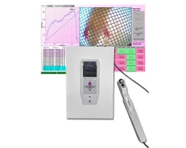
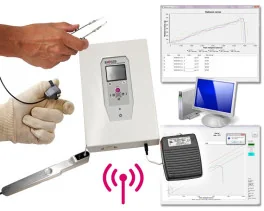
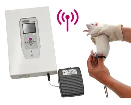
Grip strength test (BIO-GS4)
Source :