 Activity, Motor Control & Coordination
 Pain - Thermal Allodynia / Hyperalgesia
 Pain - Spontaneous Pain - Postural Deficit
 Pain - Mechanical Allodynia / Hyperalgesia
 Anxiety & Depression Disorder
 Learning/Memory - Attention - Addiction
 Physiology & Respiratory Research
 Metabolism & Nutrition
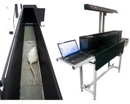
![Dynamic Weight Bearing 2.0 – Postural Module [Add-on]](https://bioseb.com/733-home_default/dynamic-weight-bearing-20-add-on-postural-module.jpg)





 Pain
Pain Central Nervous System (CNS)
 Neurodegeneration
 Sensory system
 Motor control
 Mood Disorders
 Other disorders
 Muscular system
 Joints
 Metabolism
 Cross-disciplinary subjects
 CONFERENCES & MEETINGS 2026
 Authors
Mohankumar B. Senthilkumar, Sanya Sharma, Navaneeth Srinivasan, Anila Varghese, Pratiksha Sarangi, Vijayata Singh, Narendra Kumar, Devyani Yenurkar, Sudip Mukherjee, Sonal Amit, Sameer Bhatia, Santosh K. Misra, Ratna Dua Puri, Jeffrey Chamberlain, Giridhara R. Jayandharan
Lab
Journal
Journal of Cellular and Molecular Medicine
Abstract
To corroborate the functional rescue, we performed immunostaining of different skeletal muscles, 62 weeks after gene therapy (Figure6A). A quantification of dystrophin-positive fibres in TA muscle (Mean: 50.59% vs 2.35%,p< 0.0001), GAS muscle (Mean: 59.31% vs 1.26%,p< 0.0001), and DIA muscle (Mean: 52.13% vs 3.45%,p< 0.0001) demonstrated higher dystrophin-positive fibres when compared to mock-treated mice (Figure6B). The GAS muscle demonstrates individual myofibers positive for dystrophin separated by an interstitium layer (FigureS11). The DGC proteins were prominently detected in TA muscle sections of vector–treated mice (FigureS12). The improvement in cardiac function is due to the robust expression of dystrophin in the cardiac tissue (Mean: 38.23% vs 0.05%,p< 0.0001) (FigureS13A,B). Further MT staining showed a significant reduction in collagen deposition in TA (~2.7-fold,p< 0.0001), GAS (~2-fold,p< 0.0001), and cardiac muscle (~4.24-fold,p< 0.0001) of vector-treated mice (FigureS14A,B). H&E staining also demonstrated a decrease in central nucleation of myofibers (Figure7A,B), signifying the therapeutic benefit of optimised AAV9K51Q-CoμDys. A biodistribution analysis revealed that the vectors were present in different organs such as brain, heart, lungs, kidney, liver and in skeletal muscles (TA, GAS, soleus, extensor digitorum longus (EDL), and quadriceps (QD)) (FigureS15). Interestingly, no significant changes in B cell and T cell populations were noted in treated vs control animals (FigureS16A–C), and further assessment of capsid-specific T cell response using ELISPOT assay revealed significantly reduced interferon-γ secretion in the spleenocytes of vector-treated mice (Mock vs AAV9K51Q-CoμDys, Mean: 122 vs 37 spots,p< 0.05) (FigureS16D). These data highlight the safety of the AAV9K51Q-CoμDys vectors during systemic administration. Finally, creatine kinase (CK) levels were reduced in vector-treated mice (AAV9K51Q-CoμDys vs. Mock: Mean: 478.6 U/L vs 760.7 U/L), which underscores the integrity of sarcolemma in vector-treated mice (FigureS17). Taken together, our data demonstrate the efficacy and safety of AAV9K51Q-CoμDys vector with a marked improvement in phenotype ofmdxmice, up to 1 year after vector administration.
Keywords/Topics
AAV9; codon-optimised microdystrophin; Duchenne muscular dystrophy; gene therapy
BIOSEB Instruments Used:
Grip strength test (BIO-GS4)
Source :