 Activity, Motor Control & Coordination
 Pain - Thermal Allodynia / Hyperalgesia
 Pain - Spontaneous Pain - Postural Deficit
 Pain - Mechanical Allodynia / Hyperalgesia
 Anxiety & Depression Disorder
 Learning/Memory - Attention - Addiction
 Physiology & Respiratory Research
 Metabolism & Nutrition




![Dynamic Weight Bearing 2.0 – Postural Module [Add-on]](https://bioseb.com/733-home_default/dynamic-weight-bearing-20-add-on-postural-module.jpg)









 Pain
Pain Central Nervous System (CNS)
 Neurodegeneration
 Sensory system
 Motor control
 Mood Disorders
 Other disorders
 Muscular system
 Joints
 Metabolism
 Cross-disciplinary subjects
 CONFERENCES & MEETINGS 2026
 Authors
Qiaoqi Zheng, Ji Zhuoga, Congcong Li, Wenjing Chen, Maimaititusun Yalikun, Peng Fu, Zaiquan Dong, Jingcheng Dong
Lab
Prabhjot Kaur; Morehouse School of Medicine; United States
Journal
Frontiers in Immunology
Abstract
When A549 cells were treated with COR, the expression level of PSEN1 increased (Supplementary Figure 2A), accompanied by a decrease in cell apoptosis (Supplementary Figure 2B) and an acceleration in cell proliferation (Supplementary Figure 2C). To confirm the effect of PSEN1 on lung cancer, we first established PSEN1 overexpression in A549 and H1299 cells, which was validated by western blotting (Figure 8H) and qRT-PCR (Figure 8I).In vitro, cell proliferation was enhanced following PSEN1 overexpression in both A549 and H1299, as determined by CCK8 (Figure 8J), colony formation (Figure 8K), and EdU assays (Figure 8L). Protein quantification indicated that PSEN1 was significantly upregulated in LUAD tissues compared with normal tissues (Figure 8N), a finding confirmed by immunohistochemistry (IHC) as well (Figure 8O). The high expression of PSEN1 correlated with poor overall survival in LUAD patients (Figure 8M). Next, we explored the downstream signaling pathways through which PSEN1 promotes LUAD proliferation. To investigate this, we performed enrichment analyses. Both Kyoto Encyclopedia of Genes and Genomes (KEGG,Supplementary Figure 3A) and Gene Set Enrichment Analysis (GSEA,Supplementary Figure 3B) based on TCGA-LUAD indicated an association between PSEN1 and ErbB signaling using the BEST database (https://rookieutopia.hiplot.com.cn/app_direct/BEST/). Consistently, western blotting confirmed that overexpression of PSEN1 promoted phosphorylation of ERBB1 and activation of the PI3K-AKT signaling pathway, which could be inhibited by PI3K inhibitor LY294002 (Supplementary Figure 2F). Accordingly,in vitroexperiments showed that the pro-proliferative effect of PSEN1 overexpression was attenuated by LY294002 (Supplementary Figure 2E). Furtherin vivoexperiments corroborated the tumor-promoting role of PSEN1 and demonstrated that the PI3K inhibitor Alpelisib could reverse this effect (Supplementary Figure 2G). Collectively, these results suggest that stress-induced PSEN1 may facilitate LUAD progression by activating the ERBB1-PI3K-AKT signaling pathway, a well-established driver of lung adenocarcinoma proliferation.
Keywords/Topics
cell proliferation; edu assays; luad proliferation; lung adenocarcinoma proliferation; mice; novel prognostic signature; psen1; depression-induced lung adenocarcinoma progression; a549 cells; cell apoptosis
BIOSEB Instruments Used:
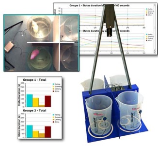
Forced Swimming Test: New FST DUAL SENSOR (BIO-FST-DSM)
Source :