 Pain - Thermal Allodynia / Hyperalgesia
 Pain - Spontaneous Pain - Postural Deficit
 Pain - Mechanical Allodynia / Hyperalgesia
 Learning/Memory - Attention - Addiction
 Physiology & Respiratory Research


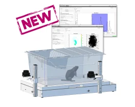

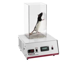
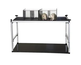
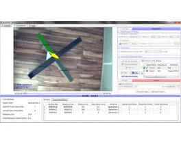
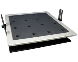
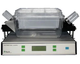
![Dynamic Weight Bearing 2.0 – Postural Module [Add-on]](https://bioseb.com/733-home_default/dynamic-weight-bearing-20-add-on-postural-module.jpg)


 Pain
Pain Central Nervous System (CNS)
 Neurodegeneration
 Sensory system
 Motor control
 Mood Disorders
 Other disorders
 Muscular system
 Joints
 Metabolism
 Cross-disciplinary subjects
 CONFERENCES & MEETINGS 2026
 Authors
Lab
Journal
Abstract
It is reasonable to assume that for neurons and their axons, lipidated peptides should readily access the membranes of unmyelinated neurons, while access to the axons of myelinated neurons would be restrictive. To test this notion, we injected 100 µM of lipidated hemagglutinin peptide (HA), a proxy for lipidated peptide distribution,25into the sciatic nerve. After 24 hours of injection, the sciatic nerve was collected and stained for peripherin, a marker for C fibers10and with an anti-HA antibody (Figs. 3A and B). The HA peptide colocalized in the peripherin-stained fibers (Pearson coefficient 0.72), indicating that the peptide preferentially partitions into the membranes of the unmyelinated C fibers and diffuses within the membrane. The HA peptide immunoreactivity is visible in the sciatic nerve up to 21 days after injection before dissipating by day 28 (Supplemental Figs. 5A and B,https://links.lww.com/PAIN/C174). There was less colocalization in NF200 positive fibers (Supplemental Fig. 5C,https://links.lww.com/PAIN/C174). Because these data demonstrated a C-fiber specific effect, we sought to determine if the PY(A) peptide would affect C-fiber activity using ex vivo skin-nerve recordings (Fig. 3C). First, hind paws of naïve mice were unilaterally injected with 20 µL of either 100 µM PY(A) peptide or scrambled peptide control. No difference in heat thermal paw withdrawal latency was observed in scrambled or PY(A) peptide–treated mice (Supplemental Fig. 6A,https://links.lww.com/PAIN/C174) indicative of intact reflexive behavior. Subsequently, the sural nerve with the skin of the hind paw was dissected from these mice for C-fiber recordings (Fig. 3B). C-fiber responses under von Frey mechanical stimulation of the receptive field did not differ between PY(A) peptide and scrambled groups (Fig. 3C). The PY(A) peptide significantly increased the electrical stimulation thresholds (Fig. 3D) and also decreased the net number of action potentials in response to heat stimulation (Figs. 3E–G). The peptide did not alter CV of C fibers in naïve mice (Supplemental Fig. 6B,https://links.lww.com/PAIN/C174) suggesting that after 24 hours, the peptide was mostly confined to the peripheral terminals. These data indicated that the PY(A) peptide blocked the sensitivity of C fibers to noxious heat stimulation in the uninjured state.
Keywords/Topics
Peptides; Neuropathic pain; Sodium channels; Scaffold protein; Trafficking; Ubiquitination; Dorsal root ganglion neurons; Electrophysiology
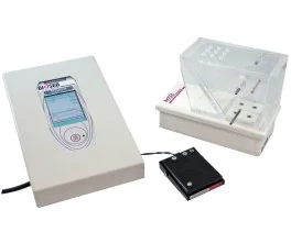
BIOSEB Instruments Used:
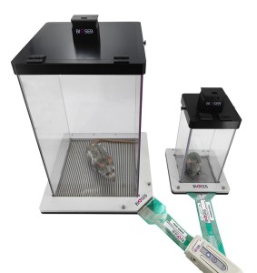
Dynamic Weight Bearing 2.0 (BIO-DWB-DUAL)
Source :