
 Activity, Motor Control & Coordination
 Pain - Thermal Allodynia / Hyperalgesia
 Pain - Spontaneous Pain - Postural Deficit
 Pain - Mechanical Allodynia / Hyperalgesia
 Anxiety & Depression Disorder
 Learning/Memory - Attention - Addiction
 Physiology & Respiratory Research
 Metabolism & Nutrition



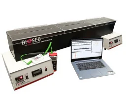
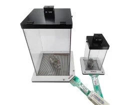
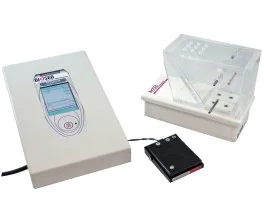
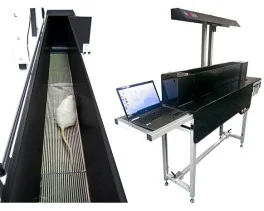
![Dynamic Weight Bearing 2.0 – Postural Module [Add-on]](https://bioseb.com/733-home_default/dynamic-weight-bearing-20-add-on-postural-module.jpg)






 Pain
Pain Central Nervous System (CNS)
 Neurodegeneration
 Sensory system
 Motor control
 Mood Disorders
 Other disorders
 Muscular system
 Joints
 Metabolism
 Cross-disciplinary subjects
 CONFERENCES & MEETINGS 2026
 Authors
Souhail Djebari, Ana Contreras, Victor Castro-Andrés, Raquel Jimenez-Herrera, Guillermo Iborra-Lázaro, Raudel Sánchez-Campusano, Lydia Jiménez-Díaz, Juan D. Navarro-López
Lab
Journal
The Journal of Physiology
Abstract
Subsequently, to explore the role of PPC functionality in this process, we surgically implanted subdural electrodes in the PPC to record LFPs in alert mice. Oscillatory activity of neural networks arises from the activity of different neuronal populations, and their synchronisation defines the amplitude (or spectral power) of oscillations at different frequency bands. In this context, GABAergic inhibitory interneurons play a pivotal role in co-ordinating the activity and synchronisation of excitatory neurons and other interneurons, thereby generating oscillations across different frequency bands (Palop & Mucke,2016). Such oscillatory activity is ultimately linked to spatial memory, changing during both navigation (Chrastil et al.,2022) and retrieval of spatial memories (Vivekananda et al.,2021). To avoid disrupting the integrity of behavioural test results, LFP recordings in this study were performed following the behavioural tests, leaving the influence of test conduct on oscillatory activity outside the scope of our examination. However, given that dysfunction within neuronal networks manifests as alterations of oscillatory activity across different frequency intervals associated with cognitive processes (Palop & Mucke,2016; Schnitzler & Gross,2005), we aimed to assess the impact of oAβ1-42i.c.v.administration on PPC's oscillatory activity. Our LFP recordings revealed that, whereas oscillatory activity in the PPC remained unaltered 24 h after oAβ1-42i.c.v.injection, a significative increase in spectral power across all analysed rhythms emerged at later time points. This effect became evident at 3 days post-i.c.v.injection in lower-frequency rhythms (δ, θ and β) and was subsequently extended to higher-frequency bands (low and high γ) by 12 days post-i.c.v.injection. This change was particularly pronounced within the θ rhythm, where the heightened spectral power enabled us to conduct a more comprehensive temporal analysis. This analysis unveiled an aberrant increase in spectral power within the θ band, starting from the third day post-injection in amyloidosis-affected animals. Consistent with our observations, alterations in θ and γ rhythms have been documented in electroencephalogram (EEG) recordings of AD patients (Adler et al.,2003; Herrmann & Demiralp,2005) and in LFP recordings of murine models of the disease (Stoiljkovic et al.,2019; Wang et al.,2020). This supports the notion that accurate memory encoding relies on these specific rhythms within task- relevant neural networks (Palop & Mucke,2016). Notably, θ and γ oscillations interact to create the θ–γ neural code, involved in sensory processing, memory and learning (Lisman & Jensen,2013), a coupling potentially disrupted in the early stages of AD (Zhang et al.,2016). Although this study did not directly address θ–γ coupling, preliminary findings based on cross-frequency comodulation suggest no significant couplings between slow and fast oscillations in our model, with somewhat stronger comodulations noted among low-frequency rhythms instead. Nevertheless, more in-depth analysis in this direction presents a compelling pathway for future research. Additionally, EEG recordings from patients in the preclinical stages of AD have also revealed alterations in other cortical rhythms, including δ and β rhythms, which may contribute to an increased risk of future cognitive decline (Gaubert et al.,2019). In addition, these findings align with other studies that describe how neuronal hyperactivity induced by Aβ in the early stages of AD and in murine models of amyloidosis can contribute to the dysfunction of neural networks and trigger epileptiform activity (Jeremic et al.,2021; Zott et al.,2018).
Keywords/Topics
Alzheimer's disease; amyloid-β (Aβ); hippocampus; LTP; oligomers; oscillatory activity; posterior parietal cortex; spatial memory
BIOSEB Instruments Used:
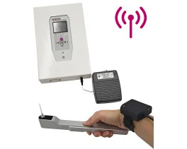
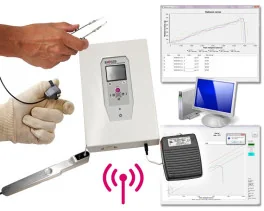
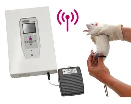
Tail Suspension Test - Wireless (BIO-TST5)
Source :
https://physoc.onlinelibrary.wiley.com/doi/abs/10.1113/JP286196