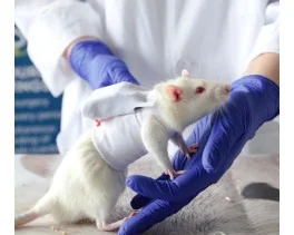
 Pain - Thermal Allodynia / Hyperalgesia
 Pain - Spontaneous Pain - Postural Deficit
 Pain - Mechanical Allodynia / Hyperalgesia
 Learning/Memory - Attention - Addiction
 Physiology & Respiratory Research
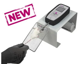
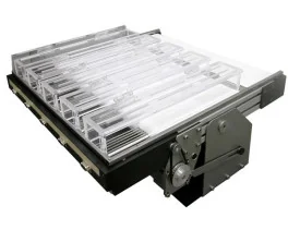

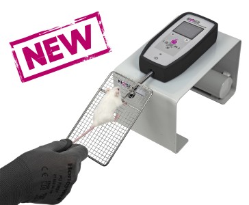
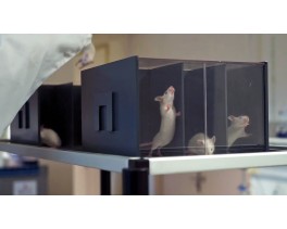
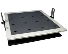
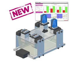
![Dynamic Weight Bearing 2.0 – Postural Module [Add-on]](https://bioseb.com/733-home_default/dynamic-weight-bearing-20-add-on-postural-module.jpg)


 Pain
Pain Central Nervous System (CNS)
 Neurodegeneration
 Sensory system
 Motor control
 Mood Disorders
 Other disorders
 Muscular system
 Joints
 Metabolism
 Cross-disciplinary subjects
 CONFERENCES & MEETINGS 2026
 Authors
Ottavia Agrifoglio, Solvig Görs, Quentin Sciascia, Zeyang Li, Elke Albrecht, Sophie Achilles, Meike Statz, Manuela Bastian, Tobias Lindner, Karen Friederike Gauß, Sarah Rohde, Karen Rischmüller, Peggy Berlin, Georg Lamprecht, Robert Jaster, Cornelia C. Metges, Luise Ehlers
Lab
Journal
Journal of Cachexia, Sarcopenia and Muscle
Abstract
In this study, we focused on early changes in metabolism and muscle function associated with CLD using the model of BDL in mice. Liver histology and basic laboratory findings indicated that BDL caused a severe cholestatic liver injury. CLD progressed over the investigation period of 14 days to a stage with liver cell necroses, inflammation, and beginning fibrosis but still without a generalized failure of liver synthesis functions as a reduced rate of hepatic protein synthesis only becomes apparent 14 days after surgery. Progressive body weight loss, lower intake of water and feed, a lower N-BAL, and reduced TEE pointed to a catabolic state of BDL mice. The lower TEE value could be explained by the lower energy requirement to metabolize the lower amount of feed ingested and by the lower energy requirement to maintain and move a lighter body [28]. However, after surgery, TEE decreased by about 20% in the BDL mice, which was related to the approximately 20% body weight loss on d3 and d10, but not to the 50% decrease in feed intake. Given that resting energy expenditure is the parameter that mostly affects TEE and that muscle energy expenditure is the primary contributor to resting energy expenditure [22], it is plausible that the weight loss of BDL mice is primarily a loss of metabolically active tissue, such as skeletal muscle–which was also detected by MRI. Interestingly, while feed and, therefore, protein intake was reduced by approximately 50% of the intake prior to BDL surgery, N-BAL was reduced by 87% at d10 compared to the N-BAL observed in healthy mice before surgery. The lower N-BAL is likely related to a disproportionately greater N loss caused by the enhanced protein catabolism associated with inflammation in liver disease [29], as suggested by the increased plasma levels of IL-6Rα, TNFRI, and TNFRII in BDL mice. The reduced feed/protein intake and the pro-inflammatory state of the BDL mice are likely to be major contributors to the reduced rates of protein synthesis in the liver and skeletal muscle, potentially exacerbating the lower N-BAL.
Keywords/Topics
cholestatic liver disease; malnutrition; metabolism; mouse model; sarcopenia
BIOSEB Instruments Used:
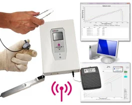
Grip strength test (BIO-GS4)
Source :