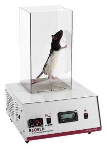
 Activity, Motor Control & Coordination
 Pain - Thermal Allodynia / Hyperalgesia
 Pain - Spontaneous Pain - Postural Deficit
 Pain - Mechanical Allodynia / Hyperalgesia
 Anxiety & Depression Disorder
 Learning/Memory - Attention - Addiction
 Physiology & Respiratory Research
 Metabolism & Nutrition
![Dynamic Weight Bearing 2.0 – Postural Module [Add-on]](https://bioseb.com/733-home_default/dynamic-weight-bearing-20-add-on-postural-module.jpg)


 Pain
Pain Central Nervous System (CNS)
 Neurodegeneration
 Sensory system
 Motor control
 Mood Disorders
 Other disorders
 Muscular system
 Joints
 Metabolism
 Cross-disciplinary subjects
 CONFERENCES & MEETINGS 2026
 Authors
Jin Ke, Wei-Cheng Lu, Hai-Yang Jing, Shen Qian, Sun-Wook Moon, Guang-Fu Cui, Wei-Xin Qian, Xiao-Jing Che, Qian Zhang, Shi-Shi Lai, Ling Zhang, Ying-Jie Zhu, Jing-Dun Xie, Tian-Wen Huang
Lab
Journal
Zoological Research
Abstract
Subsequently, we examined the function of lPBNNK1Rneurons in driving sustained pain-induced behavioral responses. Previous studies have shown that chemogenetic silencing of lPBNNK1Rneurons reduces second-phase licking elicited by hind paw formalin injection or paw clipping (Barik et al., 2021;Deng et al., 2020). To further clarify the function of lPBNNK1Rneurons, we injected AAV9-hSyn-DIO-taCasp3-TEVp virus into the lPBN ofNK1R-CreGFPmice to selectively deplete lPBNNK1Rneurons (Figure 3A, B). Mice with lPBNNK1Rneuron-ablation exhibited behavioral phenotypes similar to those observed in whole lPBN-lesioned animals. They maintained normal sensorimotor coordination (Figure 3C) and normal reflexive withdrawal responses to external mechanical and thermal stimulation (Figure 3D-H), but a marked reduction in persistent licking behavior in response to sustained 50C hot plate exposure, skin pinching, and toe clipping (Figure 3I-K). Similar to the whole lPBN-lesioned mice, the lPBNNK1R-ablated mice showed an approximate 50% reduction in the number of writhes following intraperitoneal injection of acetic acid (Figure 3L). These findings suggest a broad involvement of lPBN neurons in inflammatory irritant-evoked peritoneovisceral pain. Given that sustained intense stimulation applied to the skin or underlying deep tissues produces pain and discomfort (Henderson et al., 2006;Huang et al., 2019;Svensson et al., 1997), we hypothesized that such intense stimuli from cutaneous and/or deep tissues should produce a strong negative teaching signal, prompting the animal to learn to avoid these stimuli. To test this, we performed skin pinching- and toe clipping-evoked CPA tests (Figure 3M). We first confirmed that sustained noxious mechanical stimuli applied on the hind paw skin or bone/muscle at the toe region induced strong aversive responses inC57BL/6Jmice (Figure 3N). InNK1R-CreGFPmice, after four training sessions, the AAV-hSyn-DIO-mCherry virus-injected control group exhibited a clear aversion to the skin pinching- and toe clipping-paired compartments, whereas the lPBNNK1R-ablated mice were insensitive to such conditioning (Figure 3O). Thus, lPBNNK1Rneurons are essential for conditioned learning and/or memory evoked by noxious stimuli that produce sustained cutaneous or deep tissue mechanical pain. To further confirm the effects of lPBNNK1Rneuronal ablation, we chemogenetically silenced lPBNNK1Rneurons. We injected AAV9-hSyn-DIO-hM4D(Gi)-mCherry and control viruses into the lPBN ofNK1R-CreGFPmice, respectively, then measured cutaneous and deep tissue pain-associated behaviors (Supplementary Figure S4A). Before the behavioral tests, we first confirmed the silencing effect of CNO application by performing electrophysiological recordings on lPBN slices. Patch clamp recordings of hM4D(Gi)-mCherry-expressing NK1R+neurons indicated a complete loss of current injection-induced action potentials after bath application of CNO (Supplementary Figure S4B). Neuronal silencing was also confirmed by investigating skin pinch- or toe clip-induced c-Fos expression in the lPBN of mice after CNO injection (Supplementary Figure S4C). Mice with acute inhibition of lPBNNK1Rneurons phenocopied the lPBNNK1R-ablated mice, showing a reduction in sustained cutaneous and deep tissue pain-induced recuperative behaviors, as well as normal reflexive withdrawal reactions to external nociceptive stimuli (Supplementary Figure S4D-N). The injection sites of taCasp3, hM4Di and mCherry control virus were validated by cryosection and microscopy (Supplementary Figure S5).
Keywords/Topics
Lateral parabrachial nucleus; Substance P receptor ; Pain affect ; Defensive reaction; Somatosensory
BIOSEB Instruments Used:
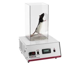
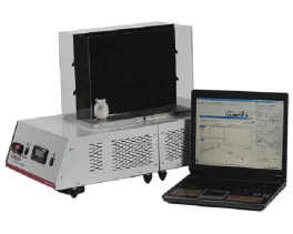
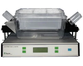
Cold Hot Plate Test (BIO-CHP)
Source :