 Pain - Thermal Allodynia / Hyperalgesia
 Pain - Spontaneous Pain - Postural Deficit
 Pain - Mechanical Allodynia / Hyperalgesia
 Learning/Memory - Attention - Addiction
 Physiology & Respiratory Research
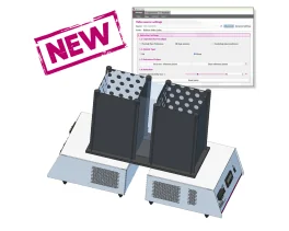
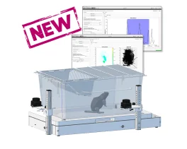
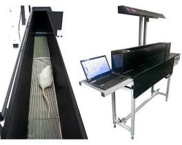
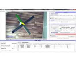
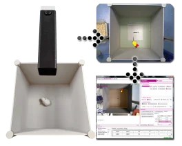
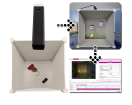
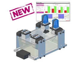
![Dynamic Weight Bearing 2.0 – Postural Module [Add-on]](https://bioseb.com/733-home_default/dynamic-weight-bearing-20-add-on-postural-module.jpg)


 Pain
Pain Central Nervous System (CNS)
 Neurodegeneration
 Sensory system
 Motor control
 Mood Disorders
 Other disorders
 Muscular system
 Joints
 Metabolism
 Cross-disciplinary subjects
 CONFERENCES & MEETINGS 2026
 Authors
Jennifer M Brazill, Ivana R Shen, Clarissa S Craft, Kristann L Magee, Jay S Park, Madelyn Lorenz, Amy Strickland, Natalie K Wee, Xiao Zhang, Alec T Beeve, Gretchen A Meyer, Jeffrey Milbrandt, Aaron DiAntonio, Erica L Scheller
Lab
Journal
JCI Insight
Abstract
The neural expression and actions of SARM1 are well established. Consistent with this, we found highSarm1expression in neural tissues, such as the DRGs (Figure 6AandSupplemental Figure 7A). By contrast,Sarm1expression by qPCR was >95% lower in bone and bone marrow (Figure 6BandSupplemental Figure 7A). Though low, this expression was still significantly reduced in Sarm1KObone relative to WT bone, with trending reductions also noted in Sarm1KObone marrow (P< 0.067). We also explored SARM1 expression at the protein level using anti-FLAG immunostaining of mouse tissues expressing 2x FLAG-tagged SARM1 (Supplemental Figure 7B). Direct immunostaining with anti-SARM1 was not possible because of poor specificity of available commercial antibodies. However, anti-FLAG immunostaining detected robust expression of FLAG-SARM1 in both DRG sensory neurons and sympathetic neurons in the aorticorenal ganglia (Supplemental Figure 7B). By contrast, FLAG-SARM1 was not detected in bone or its associated tissues using paired development procedures (analyzed cross sections of mouse bone included osteoblasts, osteoclasts, osteocytes, periosteum, adipocytes, stroma, vasculature, and bone marrow cells). This does not rule out low-level expression of SARM1 in these cells, but it reinforces prior data showing high relative expression of SARM1 in neurons (39,40). Last, we analyzedSarm1expression by qPCR in cultured bone marrow stromal cells (BMSCs), pre-osteoblasts, bone marrow-derived macrophages (BMMs) with/without LPS, and pre-osteoclasts with/without LPS from WT and Sarm1KOmice (Supplemental Figure 7, C-E). Comparable to the bulk tissue analyses, we found that low-levelSarm1expression was significantly reduced by Sarm1KOin all cell types and conditions. Future generation of extraneuralSarm1-cKO models will be needed to define the biological relevance of this low-level signal in vivo and to localize the source of the Sarm1KO-dependent skeletal protection in settings of T1D.
Keywords/Topics
Bone Biology ; Endocrinology;Bone disease ; Diabetes;Neurodegeneration In murine and diabetes and metabolic disease
BIOSEB Instruments Used:
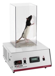
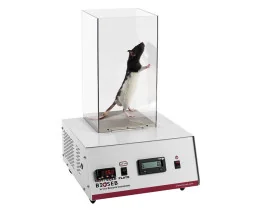
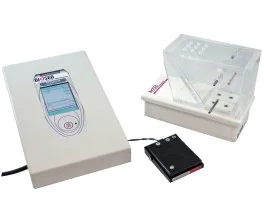
Cold Hot Plate Test (BIO-CHP)
Source :