
 Activity, Motor Control & Coordination
 Pain - Thermal Allodynia / Hyperalgesia
 Pain - Spontaneous Pain - Postural Deficit
 Pain - Mechanical Allodynia / Hyperalgesia
 Anxiety & Depression Disorder
 Learning/Memory - Attention - Addiction
 Physiology & Respiratory Research
 Metabolism & Nutrition





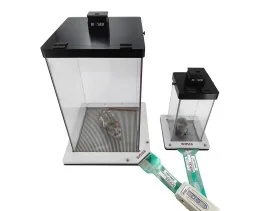
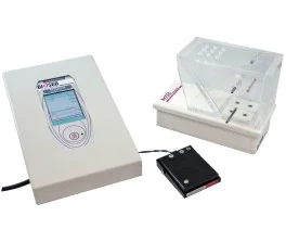

![Dynamic Weight Bearing 2.0 – Postural Module [Add-on]](https://bioseb.com/733-home_default/dynamic-weight-bearing-20-add-on-postural-module.jpg)















 Pain
Pain Central Nervous System (CNS)
 Neurodegeneration
 Sensory system
 Motor control
 Mood Disorders
 Other disorders
 Muscular system
 Joints
 Metabolism
 Cross-disciplinary subjects
 CONFERENCES & MEETINGS 2026
 Authors
Weixin Zhang, Francisco Lopez, Mei Wan, Junying Zheng, Russell Wesson, Zhaoli Sun, Xu Cao
Lab
Journal
International Journal of Surgery
Abstract
Emerging evidence identifies immune dysregulation, fibroblast-like synoviocyte (FLS) activation, and pathological neurovascular remodeling as key drivers of RA pathogenesis[9–11]. Transforming Growth Factor Beta (TGF-β) signaling plays a complex, context-dependent role, functioning as both an immunoregulatory and pathogenic factor. TGF-β1 is upregulated in the synovium of RA patients – including in synovial lining cells, macrophages, and endothelial cells – and is elevated in synovial fluid compared to osteoarthritis and healthy controls[12–14]. In RA, TGF-β promotes Th17 cell differentiation in the presence of IL-6, contributes to synovial fibrosis and hyperplasia, and enhances pathological angiogenesis[15–17]. In conjunction with inflammatory cytokines such as TNF-α, IL-1β, and IL-6, TGF-β upregulates receptor activator of NF-κB ligand (RANKL) in FLSs, promoting osteoclastogenesis and bone erosion[18,19]. The imbalance between pro-inflammatory Th17 cells and regulatory T cells (Tregs), alongside elevated TNF-α and IL-17A, further perpetuates chronic inflammation and joint destruction[8,20]. Additionally, aberrant angiogenesis and sensory nerve innervation within the synovial microenvironment exacerbate joint pain. Targeting the TGF-β pathway and its downstream mediators (e.g., SMADs) offers therapeutic potential but requires selective modulation to preserve essential immunoregulatory functions, such as Treg maintenance.
Keywords/Topics
pathological neurovascular remodeling; joint pain; surgery; immune dysregulation; fibroblast-like synoviocyte fls activation; key drivers; transforming growth factor beta; tgf-β; tgf-β1; ra patients
Source :