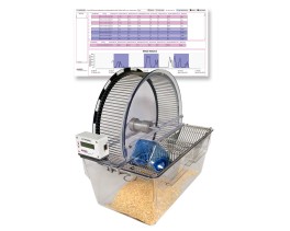
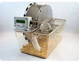
 Activity, Motor Control & Coordination
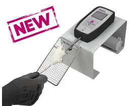
 Pain - Thermal Allodynia / Hyperalgesia
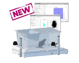
 Pain - Spontaneous Pain - Postural Deficit
 Pain - Mechanical Allodynia / Hyperalgesia
 Anxiety & Depression Disorder
 Learning/Memory - Attention - Addiction
 Physiology & Respiratory Research
 Metabolism & Nutrition
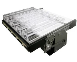
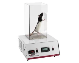
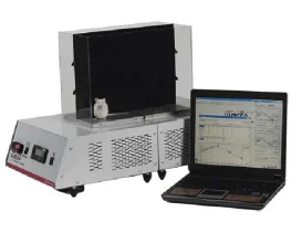
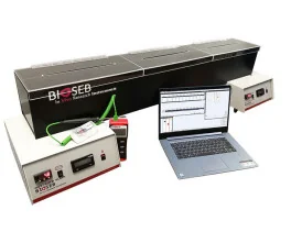





 Pain
 Central Nervous System (CNS)
 Neurodegeneration
 Sensory system
 Motor control
 Mood Disorders
 Other disorders
 Muscular system
 Joints
 Metabolism
 Cross-disciplinary subjects
 CONFERENCES & MEETINGS 2026
Authors
Irina Shchukina, Carlos J. Rodriguez-Hernandez, Heather S. Ruiz, Maksim Kleverov, Rachel L. Mintz, Katsutaka Mineura, Subhadra C. Gunawardana, Sunnie Hsiung, Bishan Bhattarai, Veronika Vachova, Jan Kossl, Denis A. Mogilenko, Rachael L. Field, Tran H. Nguyen, Quazim A. Alayo, Christopher G. Huckstep, Barbora Vander Wielen, Jonathan R. Brestoff, Sheila A. Stewart, David W. Piston, Takeshi Egawa, Daniel Kreisel, Gwendalyn J. Randolph, Maxim N. Artyomov
Lab
Journal
Cell Reports
Abstract
Summary
Aging strongly impacts CD8+ T cells, including the loss of naive cells and the emergence of age-associated GZMK+CD8+ T cells (TAA cells). Although TAA cells constitute a major population in aged mice, the pathway underlying their differentiation remains unknown. Here, we demonstrate that TAA cell development is cell extrinsic and requires antigen exposure within aged non-lymphoid tissues. Using a TNFΔ69AU/+ mouse model, we show that low-grade inflammation accelerates CD8+ T cell aging and promotes early accumulation of TAA cells. Analysis of TAA cell heterogeneity further identifies a progenitor subpopulation enriched in the aged adipose tissue. Finally, heterochronic transplantation experiments suggest that the aged adipose tissue can serve as a systemic source of TAA cells and contribute to the conversion of young CD8+ T cells into the aged phenotype. Together, these findings indicate that aged non-lymphoid tissues actively drive CD8+ T cell remodeling and identify adipose tissue as an important niche shaping immune aging.
Keywords/Topics
aging; CD8+ T cells; inflammaging; granzyme K; adipose tissue; CP: immunology
BIOSEB Instruments Used:
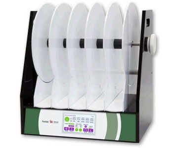
Bioseb CIS Software (BIO-CIS2),Grip strength test (BIO-GS4),Rotarod for rats and mice (BX-ROD)
Source :
https://www.cell.com/cell-reports/fulltext/S2211-1247(26)00381-5