 Pain - Thermal Allodynia / Hyperalgesia
 Pain - Spontaneous Pain - Postural Deficit
 Pain - Mechanical Allodynia / Hyperalgesia
 Learning/Memory - Attention - Addiction
 Physiology & Respiratory Research
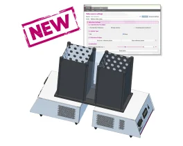
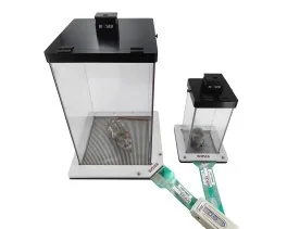
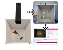
Contextual Kits for T2CT - Dual Aversion & CPP
The Contextual Kits for T2CT have been designed to enhance thermal place preference studies...
The Contextual Kits for T2CT have been designed to enhance thermal place preference studies...
An easy way to objectively quantify the muscular strength of mice and rats, and to assess the...
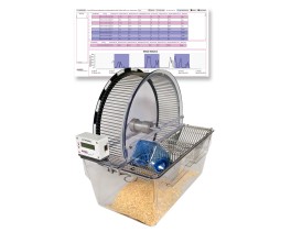
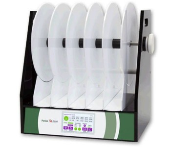
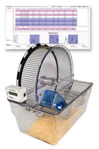
The BIOSEB Spontaneous Activity Wheel offers an effective solution for quantifying rodent...
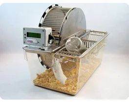
The BIOSEB Spontaneous Activity Wheel is an easy way to quantify rodent voluntary activity in...
The uncomplicated way to monitor rodent activity over several days from their home cage...

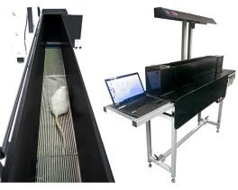
These small animal treadmills are used for forced exercise training and accurate testing of...
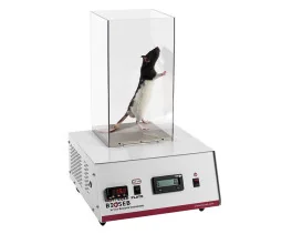
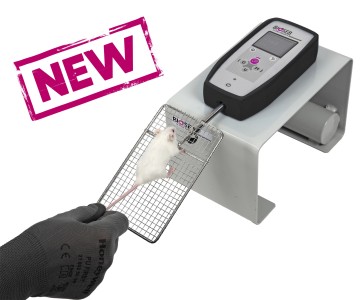
For testing animal's thermal sensitivity to pain resulting from exposure to heat or cold: the...
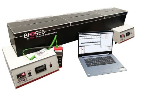
An operator independent test to study pain thresholds in rodents (mouse and rat) by assessing...
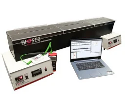
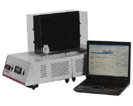
New and improved! The operator-independent Thermal Gradient Test used to show favorite...
The Contextual Kits for T2CT have been designed to enhance thermal place preference studies...
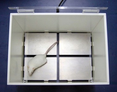
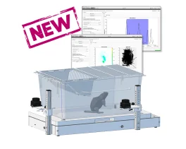
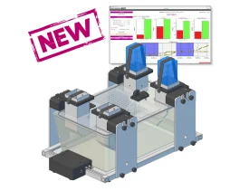
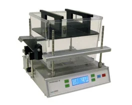
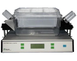
The advanced version of our Dynamic Weight Bearing Test for rodents (rats and mice) allows for...
An easy and non pain-inducing solution for assessing the level of discomfort (incapacitance) in...
A unique device that provides automated measurement of weight bearing and force distribution...
![Dynamic Weight Bearing 2.0 – Postural Module [Add-on]](https://bioseb.com/733-home_default/dynamic-weight-bearing-20-add-on-postural-module.jpg)
Expand Your Analysis with Advanced Postural and Locomotor Calculations BIOSEB’s renowned...
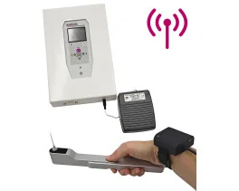
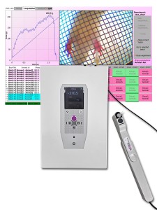
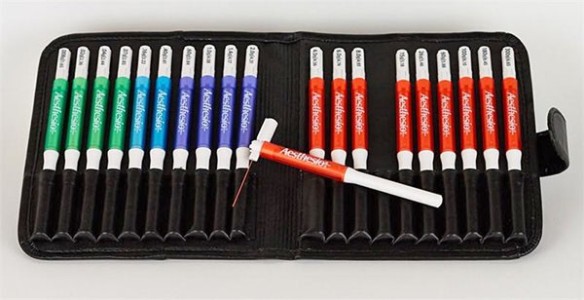
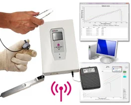
A quick solution to determine the mechanical sensitivity threshold in rodents (mice and rats)....
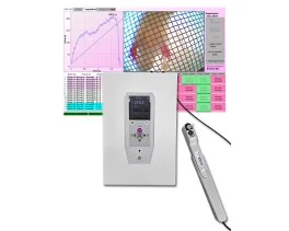
As an electronic version of the classical Von Frey Filaments esthesiometer (or aesthesiometer),...
New ROBUST and modular cages to gently hold rodents (rats or mice) during nociceptive and...
An economical and versatile solution for when flexible quantitative sensory testing (QST) is...
Dedicated to small animals, like mice and rats, Smalgo is a pressure-based analgesimeter...
Bioseb's version 5 of the Tail Suspension Test system, based on both strain sensors and video...
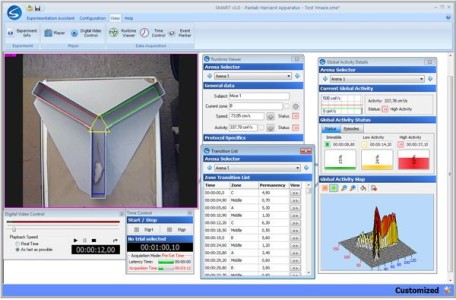
NEW ! A complete (hardware + software), dedicated and automated solution for the Elevated Plus...
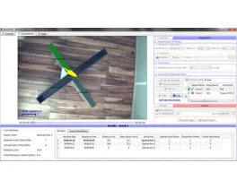
A unique setup for the automation of the Open Field test for rats and mice : 3D-camera based...

Open-field test - ARENA ONLY - used to evaluate of animal's basal activity and its evolution for...

The new Forced Swimming Test system from Bioseb uses a dual approach: Combining a double input...
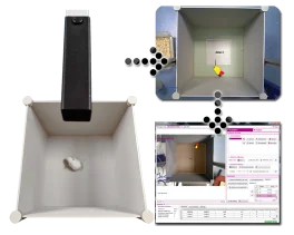
A brand new innovative setup for the automation of the Novel Object Recognition Test : 3D-camera...
Open-field test - ARENA ONLY - used to evaluate of animal's basal activity and its evolution for...

An entirely modular experimental enclosure designed to conduct operant conditioning procedures...
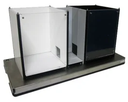
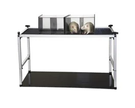
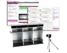
A standard experimental chamber for automated or manual assessment of conditioned place...

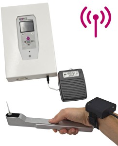
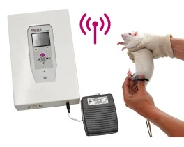
Real-Time Physiological Monitoring for Small Animals – Wireless & Non-Invasive The Bioseb...
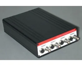
The ETH-401 is a bridge amplifier for various transducers that provides four channels of...
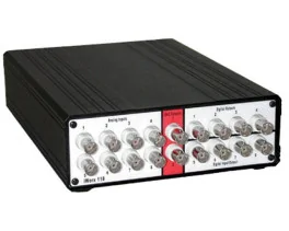
The IX-118 is a fast 100 Khz, high-resolution data acquisition system suitable for most data...
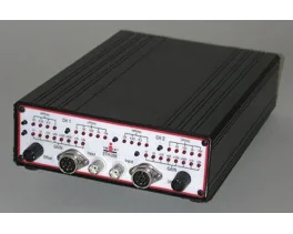
The ETH-256 is a 2 channels high performance, general-purpose life science research amplifier,...
Discover BIO-FOODIS, the next generation solution for understanding animal feeding behavior with...
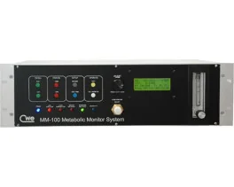
The OXYLET system - Physiocage is a modular system allowing the integration of respiratory...
Innovative and appropriate equipment for measuring food/liquid consumption and correlated motor...
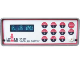
An economical, high performance Oxygen and Carbon Dioxide Analyzer with sampling rates fast...
 Latest publication 03/23/2015
 Primary coenzyme Q10 (CoQ10) deficiency is due to mutations in genes involved in CoQ biosynthesis. The disease has been associated with five major...
Read moreFilters
 Pain
Pain Central Nervous System (CNS)
 Neurodegeneration
 Sensory system
 Motor control
 Mood Disorders
 Other disorders
 Muscular system
 Joints
 Metabolism
 Cross-disciplinary subjects
 CONFERENCES & MEETINGS

 [img_empty] => /var/www/vhosts/de3310.ispfr.net/bioseb2024/modules/prestablog/views/img/product_link_white.jpg
[image_presente] => 1
[link] => https://bioseb.com/en/activity-motor-control-coordination/40-spontaneous-activity-wheels.html
)
[1875] => Array
(
[name] => Basic spontaneous activity wheels
[description_short] =>
[img_empty] => /var/www/vhosts/de3310.ispfr.net/bioseb2024/modules/prestablog/views/img/product_link_white.jpg
[image_presente] => 1
[link] => https://bioseb.com/en/activity-motor-control-coordination/40-spontaneous-activity-wheels.html
)
[1875] => Array
(
[name] => Basic spontaneous activity wheels
[description_short] => 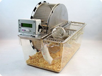 [img_empty] => /var/www/vhosts/de3310.ispfr.net/bioseb2024/modules/prestablog/views/img/product_link_white.jpg
[image_presente] => 1
[link] => https://bioseb.com/en/activity-motor-control-coordination/1875-spontaneous-activity-wheels.html
)
)
)
1
[img_empty] => /var/www/vhosts/de3310.ispfr.net/bioseb2024/modules/prestablog/views/img/product_link_white.jpg
[image_presente] => 1
[link] => https://bioseb.com/en/activity-motor-control-coordination/1875-spontaneous-activity-wheels.html
)
)
)
1